Encephalomyocarditis virus (EMCV) is small non-enveloped
ssRNA with a genome of 7.8 kb and thus considerably smaller than CoV. Being the
causative agent of myocarditis, encephalitis, reproductive disorders, diabetes,
and neurological diseases, EMCV pathogenesis is viral strain and host specific.
Although EMCV infection in humans are only associated with low morbidity, the
use porcine xenografts may cause future problems.
EMCV and the
autophagy pathway
Permissive rodent cell lines derived from hamster, rat and
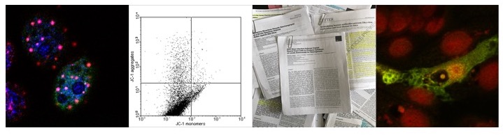
mice infected with EMCV exhibited an increase in LC3-II positive double or
single membrane vesicles in the perinuclear region indicating that the
infection of cells with EMCV induces autophagy, although in BHK-1 cells the
kinetics are slower compared to rat H9c2 or murine BV cells. Furthermore, the
induction of autophagosomes is dependent on viral replication as UV inactivated
virus fails to increase the levels of LC3-II. The formation of autophagosomes
increases over time as a cumulative amount of LC3-II increases as the infection
progresses., although the increase of LC3-II itself however only indicates that
autophagosomes are accumulating but not necessarily subject to lysosomal
degradation. Treatment of infected cells with E64d -an inhibitor of lysosomal
hydrolases- therefore should increase the number of LC3-II positive punctae, as
it indeed it is the case in EMCV infected BHK-1 cells; EMCV infection therefore
enhances the autophagic flux. Interestingly, the treatment of EMCV infected
cells also increases the levels of the viral VP1 protein, suggesting that the
induction of autophagy might be a cellular antiviral response rather than
beneficial for viral replication. Concomitant
with the increase in the formation of autophagosomes is a degradation of p62/SQSTM1,
which initially suggested that EMCV might induce selective autophagy. In the
light of recent results indicating that the Coxsackievirus B3 viral proteases
3CPro and 2APro cleave cellular p62/SQSTM1 and that the p62/SQSTM1
C-terminal fragment inhibits the formation of p62/SQSTM1 positive punctae, this
result needs to be revisited and it is
the opinion of the author that EMCV might not induce selective autophagy per se
- which of course does not rule out that one or more viral protein might do so.
Indeed, recent results suggest that the overexpression of the viral protein 2C
and 3D in the absence of the viral proteases induces the degradation of
p62/SQSTM1 by selective autophagy.
The notion that the induction of autophagy by EMCV
constitutes an antiviral response is further supported by finding that in
infected MEF treated with Bafilomycin A or siRNA against Beclin-1 or ATG5 viral
replication increases rather than decreased. This picture is however is
complicated by finding that the effect of Bafilomycin A treatment on viral
yields depends on when the virus was harvested -with viral yields higher at 7 h
p.i. in BafA1 treated compared to untreated cells whereas at 12 and 16 h p.i.
autophagy inhibition reduces the viral yield. In other words, at the early
stages of infection the increase of autophagy induces an antiviral response
whereas later in infection the formation of autophagosomes favours viral
replication. How can these conundrums being solved? When we look closer at the
experiments conducted with E64d, which suggest that the autophagosomes formed
are degraded within the lysosome then we see that cell extracts from cells at
more than 16 h p.i. were not analysed. It may therefore possible that from 16 h
p.i. onwards, the autophagosomes formed are not degraded but part of the viral
replication centers. In a further twist,
the kinetics might differ between the viral strain and the cell line used as
well as dependent on the MOI.
So if the induction of autophagy early in the replication
cycle can be considered an anti - rather than a pro-viral pathway, then we have
to ask, how is this pathway induced? Being a Picornavirus, EMCV has a ssRNA
genome. As discussed for the Coronaviridae, ssRNA viruses produce dsRNA intermediates
that alongside the viral ssRNA can be recognised by cellular pathogen
recognition receptors (PRR), which in turn induce the expression of cytokines,
including Interferon-γ (IFN-γ). Among other genes, IFN-γ induces the expression of 2,5
oligoadenylate synthetase (2,5-OAS) in a STAT1 and BRCA1 dependent manner.
2,5-OAS in turn activates RNaseL, thus degrading viral RNA and damaging
components of the host cell -including ribosomes- required for viral
replication. RNaseL has also been shown to induce autophagy and it is indeed
this mechanism which might induce autophagy early in infection by
phosphorylating Bcl-2 in N terminal JNK
dependent manner, thus dissociating BcL-2 from Beclin-1. Interestingly,
expression and activation of RNaseL also degrades p62/SQSTM1, suggesting that
at least early in infection it is RNaseL and not the viral 3CPro protease
that cleave p62/SQSTM1, which is further supported by results that in cells
expressing an inactive R667A RNaseL mutant p62/SQSTM1 is not degraded at 16 h
p.i. Although so far the precise mechanism has not been elucidated, EMCV and
other Picornaviruses have been postulated to inhibit RNaseL later in infection.
The expression of the EMCV 2C and 3D proteins has been shown to induce all three branches of the ER stress response, PERK, ATF6α and IRE1, as characterised by the phosphorylation of eIF2α, cleavage of XBP1, and increase of CHOP. Unfortunately so far the expression levels of autophagy-related genes have not been examined. If the switch coincides with an accumulation of the viral 3CPro protease is not known: as outlined above, 3CPro might cleave the MAM complex akin to Hepatitis C Virus NS3/4. Also, cleavage of the MAVS might lead to mitochondrial fission in similar way as Respiratory Syncytial Virus NS1/NS2 proteases. As it is the case in RSV infected cells, this would inhibit antiviral signalling by the MAVS/TRAF3/TRAF6 pathway, suppressing the type I interferon response and thus favour viral replication.
The induction of the ER stress response itself is probably a result of lipid repletion induced by the excessive formation of membranous vesicles - not only by viral proteins directly but also the induction of autophagy by RNaseL and indeed the induction of MAM induces the transfer of lipids to Mitochondria.
In any case, the activation of RNaseL via 2,5-OAS binding to viral RNA not only degrades viral RNA and inhibits viral replication, but also allows the recognition of these RNA fragments by PACT and MDA5. Both proteins bind to the mitochondrial-associated adaptor molecule MAVS and thus activating a pathway which ultimately increases the expression of IFN-β and other chemokines. In addition - and this is the important point in the context discussed in this post- PPRs such as RIG-1 or the RIG-1 like protein MDA5 both of which contain a MAVS domain also bind to the ER where they form a mitochondrial-associated membrane (MAM) complex via MAVS, forming an immune synapse. Recruitment of ATG14L via Syntaxin 17 and ATG5 initiates then the formation of autophagosomes and the degradation of viral RNA via autophagy. Consequently, in the context of EMCV, during the early stages of the infection cycle, the viral RNA not only activates the RNaseL system, but also indirectly increases autophagy. If this requires the MAVS signalling pathway and the inhibition of this pathway increases viral yield at early timepoints remains to be seen. In the case of Hepatitis C Virus, the viral NS3/4A protease cleaves MAVS and prevents the formation of MAM complexes by targeting the synapse. In the case of EMCV, the viral 3CPro protease might target the synapse as well - only later in infection.
 |
| Induction of 2,5OAS/RNaseL signalling and MAM by EMCV viral RNA induces autophagy |
In relation to the potential of inducing the formation of LC3 positive vesicles, a number of viral proteins have been shown to co-localise with LC3 in vesicular structures, in particular VP1, VP4, 2B, 3A, 2C, and 3D. With the exception of the viral 2C and 3D proteins however none of them induces the formation of autophagosomes as determined by the conversion of LC3-I to LC3-II, suggesting that LC3-I positive vesicles containing these proteins might play a role as transport vesicles. The question remains however, if the expression of these proteins does inhibit the formation of autophagosomes under conditions of starvation or amino-acid withdrawal. Also, it might be interesting to see whether these vesicles are positive for LC3-C, analogous to LC3-C positive vesicles containing the Capsid protein of Chikungunya Virus. Although it has been postulated that EMCV replication takes place on LC3 positive membranes, it is not clear if these represent mature autophagosomes - which is rather unlikely, unless fusion with the lysosome is prevented or if these “replication vesicles” fuse with components of the endosomal system instead. In the case of the viral 2C and 3D proteins, the formation of autophagosomes and subsequent fusion with lysosomes seems not to be inhibited, a process that also degrades p62/SQSTM1; indeed, autophagosomes containing 2C and 3D are positive for p62/SQSTM1 and induce the degradation of the latter. Presently, it is not clear however if p62/SQSTM1 is required for 2C and 3D to undergo autophagic degradation.
 |
| 2C and 3D induce the formation of mature autophagsomes |
The expression of the EMCV 2C and 3D proteins has been shown to induce all three branches of the ER stress response, PERK, ATF6α and IRE1, as characterised by the phosphorylation of eIF2α, cleavage of XBP1, and increase of CHOP. Unfortunately so far the expression levels of autophagy-related genes have not been examined. If the switch coincides with an accumulation of the viral 3CPro protease is not known: as outlined above, 3CPro might cleave the MAM complex akin to Hepatitis C Virus NS3/4. Also, cleavage of the MAVS might lead to mitochondrial fission in similar way as Respiratory Syncytial Virus NS1/NS2 proteases. As it is the case in RSV infected cells, this would inhibit antiviral signalling by the MAVS/TRAF3/TRAF6 pathway, suppressing the type I interferon response and thus favour viral replication.
 |
| Formation of the MAM complex at the ER |
The induction of the ER stress response itself is probably a result of lipid repletion induced by the excessive formation of membranous vesicles - not only by viral proteins directly but also the induction of autophagy by RNaseL and indeed the induction of MAM induces the transfer of lipids to Mitochondria.
Consequently, the MAM complex induced by the viral RNA leads to the induction of autophagy, a process that ultimately degrades the viral RNA as well as MDA5. Inducing autophagy early in the infection therefore has an antiviral effect which in turn depletes the cell of MDA5, which combined with the inhibiton of host cell translation would create favourable conditions for viral replication. Indeed, in murine L cells infected with EMCV, host cell translation decreases by 50% within the first 6 h p.i. .
 |
| Connecting the dots:RNaseL activation induces the formation of the Mitoxosome and subsequent mitophagy |
However, since only a relative small number of studies addressed the formation of the replication complexes of EMCV directly, the details of the induction of autophagosomes or autophagosome like structures during EMCV infection are not very well characterised. What emerges however, is a picture where EMCV similar to other RNA viruses hijacks the autophagic pathway in order to facilitate the formation of replication centers or replication compartments. One question which needs to be answered is the role of EMCV 2C in inhibiting the viral protease early in the infection cycle and thus preventing the degradation of RIG-1. If this is the case, then expressing 2C in trans might prevent autophagy induced by RNaseL and increase virus yield. Finally, is the induction of autophagy in EMCV infected cells by viral proteins required to prevent the induction of apoptosis which would be expected following the activation of RNaseL? One question I personally would like to address is if the infection of cells with EMCV re-localises mitochondria to MAM and thus induces mitophagy.
Targeting mitochondria dependent antiviral signalling however is not confined to cells infected with EMCV or Hepatitis C Virus. In the case of SARS-CoV, the viral orf9b protein suppresses MAVS dependent signalling and induces ATG5 dependent mitophagy, suggesting again that different viruses subvert common pathways.
As for JEV, Coxsackievirus B3, or CoV replication, the ER stress response is central for EMCV replication. Personally, the author of these lines hopes to contribute to solving some of the puzzles.
Further reading
Carocci, M., & Bakkali-Kassimi, L. (2012). The encephalomyocarditis virus Virulence, 3 (4), 351-367 DOI: 10.4161/viru.20573
Hou, L., Ge, X., Xin, L., Zhou, L., Guo, X., & Yang, H. (2014). Nonstructural proteins 2C and 3D are involved in autophagy as induced by the encephalomyocarditis virus Virology Journal, 11 (1) DOI: 10.1186/1743-422X-11-156
Zhang Y, Li Z, Ge X, Guo X, & Yang H (2011). Autophagy promotes the replication of encephalomyocarditis virus in host cells. Autophagy, 7 (6), 613-28 PMID: 21460631
Shi J, Fung G, Piesik P, Zhang J, & Luo H (2014). Dominant-negative function of the C-terminal fragments of NBR1 and SQSTM1 generated during enteroviral infection. Cell death and differentiation, 21 (9), 1432-41 PMID: 24769734
Maurel M, Chevet E, Tavernier J, & Gerlo S (2014). Getting RIDD of RNA: IRE1 in cell fate regulation. Trends in biochemical sciences, 39 (5), 245-54 PMID: 24657016
Silverman RH (2007). Viral encounters with 2',5'-oligoadenylate synthetase and RNase L during the interferon antiviral response. Journal of virology, 81 (23), 12720-9 PMID: 17804500
Siddiqui MA, & Malathi K (2012). RNase L induces autophagy via c-Jun N-terminal kinase and double-stranded RNA-dependent protein kinase signaling pathways. The Journal of biological chemistry, 287 (52), 43651-64 PMID: 23109342
Chakrabarti A, Ghosh PK, Banerjee S, Gaughan C, & Silverman RH (2012). RNase L triggers autophagy in response to viral infections. Journal of virology, 86 (20), 11311-21 PMID: 22875977
Mullan PB, Hosey AM, Buckley NE, Quinn JE, Kennedy RD, Johnston PG, & Harkin DP (2005). The 2,5 oligoadenylate synthetase/RNaseL pathway is a novel effector of BRCA1- and interferon-gamma-mediated apoptosis. Oncogene, 24 (35), 5492-501 PMID: 15940267
Hamasaki M, Furuta N, Matsuda A, Nezu A, Yamamoto A, Fujita N, Oomori H, Noda T, Haraguchi T, Hiraoka Y, Amano A, & Yoshimori T (2013). Autophagosomes form at ER-mitochondria contact sites. Nature, 495 (7441), 389-93 PMID: 23455425
Jen G, & Thach RE (1982). Inhibition of host translation in encephalomyocarditis virus-infected L cells: a novel mechanism. Journal of virology, 43 (1), 250-61 PMID: 6287000
Banerjee R, Weidman MK, Echeverri A, Kundu P, & Dasgupta A (2004). Regulation of poliovirus 3C protease by the 2C polypeptide. Journal of virology, 78 (17), 9243-56 PMID: 15308719
Shi CS, Qi HY, Boularan C, Huang NN, Abu-Asab M, Shelhamer JH, & Kehrl JH (2014). SARS-Coronavirus Open Reading Frame-9b Suppresses Innate Immunity by Targeting Mitochondria and the MAVS/TRAF3/TRAF6 Signalosome. Journal of immunology (Baltimore, Md. : 1950) PMID: 25135833
No comments:
Post a Comment